
The Blueprint We Never Drew
We do not have an imaging problem. We have a design problem.
Origins
Radiology traces its origins to 1895, when Wilhelm Röntgen was alone in his laboratory in Würzburg, Germany, experimenting with a cathode ray tube—an early glass apparatus used to study electricity. To block out visible light, he wrapped the tube in black cardboard and darkened the room. When he switched the device on, a small fluorescent screen across the room began to glow.1
This should not have been possible. The tube was covered. Light could not escape. Whatever was emanating from the apparatus was invisible—and capable of passing through solid objects. Röntgen had discovered what he called a "new kind of ray."1
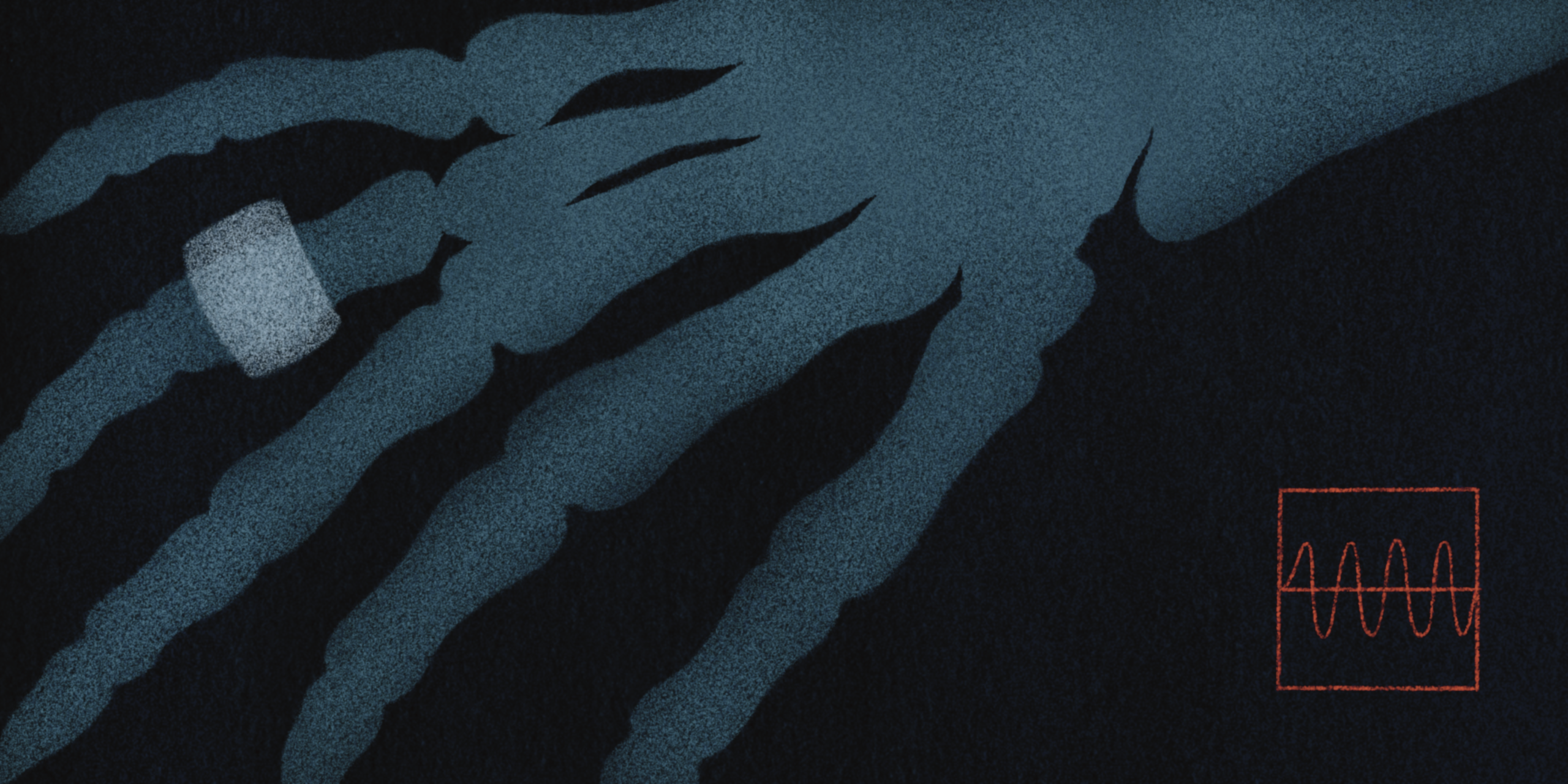
In trying to understand what he was seeing, he asked his wife, Anna, to place her hand between the tube and the screen. The image appeared almost instantly. Her bones were clearly visible, as was the outline of her wedding ring. It was the first time the interior of the human body had been seen without incision. Anna Röntgen looked at the image and said she was seeing her own death. She was not wrong to be unsettled. There was her hand, stripped of everything but bone and metal. What she could not have known was that within a generation, this process would be ordinary. And within a century, medicine could not function without it. Röntgen named the phenomenon "X-rays," the "X" standing for the unknown, capturing both mystery and possibility.3
The discovery spread with astonishing speed. Within weeks, the experiment was reproduced across Europe and North America. In 1896, The Lancet initially dismissed the findings, only to concede days later that the discovery would "produce quite a revolution in the examination of the interior of the human body."2 Hospitals soon acquired X-ray machines, and the earliest radiology departments took shape. Military applications followed almost immediately.3
As new imaging modalities emerged, diagnostic capability expanded dramatically. Fluoroscopy enabled real-time visualization. By the latter half of the 20th century, CT, MRI, and PET transformed diagnosis, staging, and treatment planning across nearly every specialty.3 What began as a scientific curiosity became indispensable. Imaging was no longer optional. It had become an imperative of modern medicine.
Before 1895, medicine was largely a solitary pursuit. Physicians diagnosed illness, set fractures, and delivered babies using little more than clinical judgment and their own hands. Consultation existed, but it was optional. X-rays changed that permanently. Good medicine now required specialized equipment and physicians whose sole expertise was image interpretation. Diagnosis became a shared act. For the first time at scale, medicine became truly interdependent, and it happened faster than the system could adapt.3
What Röntgen could not have imagined was the coordination required to integrate imaging seamlessly into patient care. Technology advanced rapidly, but the systems needed to manage it did not keep pace. We built extraordinary imaging capability without designing the infrastructure required to govern it.
That omission still shapes our experience of care today.
The Bottleneck
As reliance on diagnostic imaging increased, referral volumes rose sharply. Demand driven by aging populations, broadening indications, and rising patient expectations began to outstrip capacity. Few structural limits were placed on ordering. Radiology was often perceived as an ordering service rather than a consultation service.4
The consequences were predictable. Imaging systems became burdened by inefficiencies and overuse, including a substantial proportion of low-value imaging, such as studies ordered for inappropriate indications or ones that added little to patient management.5
No engineering team would accept a system in which unlimited demand enters through fragmented channels, urgency is inconsistently defined, work remains largely invisible until a crisis occurs, and success is measured primarily by volume. Yet this is effectively how diagnostic imaging operates across much of Canada.6
Appropriate and inappropriate requests compete for the same limited resource, and the system lacks a consistent way to tell them apart. Urgency is self-declared rather than standardized, with "urgent" designations applied variably, and often without sufficient justification.4
Some imaging studies genuinely require rapid access. Many others, however, are advanced to work around bottlenecks, manage patient pressure, or avoid the risk of delay, not because of true clinical need.5
The pathways for expediting care are poorly defined, leaving clinicians uncertain about how to access timely imaging when it truly matters. In the absence of better signals, wait times become the dominant metric. They measure everything and explain nothing.7
The true impact of uncoordinated demand is best understood through two patients who entered the same imaging queue in 2019.
The first was a 55-year-old accountant with intermittent, nonspecific abdominal pain. He had felt it on and off for a few weeks—nothing severe enough to stop him from working or sleeping. Bloodwork and physical examination were entirely normal. There was no weight loss, anaemia, or concerning history. His physician reassured him that no investigations were needed, and they agreed on a plan for watchful waiting. However, his wife had been to the emergency department the previous year with abdominal pain. She was ultimately diagnosed with diverticulitis, spent several days on intravenous antibiotics, and recovered. The experience had shaken them both. Now, every time her husband mentioned his stomach, she saw herself in the ER. She wanted him to get a CT scan. He wanted to be scanned. "Just to be safe," he told his doctor at the follow-up appointment. And again at the next one.
Eventually, the referral was sent. The scan revealed multiple incidental findings: small renal and hepatic cysts, mild diverticulosis, and degenerative changes in the lumbar spine. None explained his pain, which by that point had already resolved. What followed was the familiar and unnecessary cascade: follow-up imaging to monitor the cysts, referrals to urology and gastroenterology, and growing anxiety over findings that were never clinically significant. Six months after the initial scan, his abdominal discomfort was gone—but he was checking his phone daily for specialist appointment confirmations. Capacity was consumed. Outcomes were unchanged.
The second patient, presenting to the same clinic, was a 75-year-old retired teacher who lived alone. She had diabetes and presented with worsening abdominal pain and early satiety. She had lost twelve pounds without trying. She felt full after just a few bites of food. She looked tired in a way that worried the doctor.
Bloodwork showed anaemia. The requisition for a CT scan was marked routine, with a note: "rule out malignancy." The estimated wait time was twelve weeks.
At eighteen weeks, the patient called the clinic. No scan yet. The pain had worsened enough that she went to the emergency department. CT imaging obtained overnight revealed an obstructing colon cancer with early metastatic disease. Surgical review documented that earlier detection may have enabled curative resection prior to obstruction and metastatic spread. The patient required urgent diversion and subsequent systemic therapy.
She survived, but with more advanced disease and significantly greater morbidity than would likely have occurred with timely imaging.
These patients never met. They had different doctors, different lives, different outcomes. But they shared the same queue. The first patient's scan, and thousands like it, consumed capacity that the second patient needed. The system processed both as "demand" and distinguished neither by urgency nor appropriateness. No individual decided to prioritise one over the other. The system simply could not tell the difference.
In systems with finite capacity, low-value care inevitably displaces high-value care. The victims are often invisible. Patients wait longer, grow more anxious, and undergo cascades of further testing triggered by incidental findings. Downstream services absorb the cost. When imaging stalls, the system stalls with it.7 As Berwick observed, every system is perfectly designed to get the results it gets.6 The current state of imaging access should not surprise us.
The problem is structural. When pressure builds, the reflex response is to add capacity: more scanners, extended hours, faster throughput. These measures offer only temporary relief. Without coordinated demand, reduced duplication, and evidence-based pathways, any gains are quickly absorbed.
When access to outpatient imaging falters, patients adapt. Many seek alternate entry points into the system. Conditions that might have been managed electively present urgently after weeks or months of delay. Those with financial means may pursue private imaging, but this does not resolve the underlying problem: incidental findings still require follow-up and often publicly funded downstream care, placing further burden on the already constrained system. For vulnerable patients, delays can mean prolonged hospitalisation, loss of independence, financial hardship, and even death.
Urgent presentation reshapes care in predictable ways. Add-on imaging displaces scheduled work. Guidelines and decision support are bypassed. Clinical histories are abbreviated. Imaging becomes defensive rather than deliberate, serving as a tool for triage and risk transfer rather than thoughtful diagnosis. Care that could have been planned is instead delivered in the most expensive settings. The same scan costs more when staffing is stretched, workflows are disrupted, and competing priorities dominate. The result is not better care, but more waste.
We do not have an imaging problem. We have a design problem. Until demand is coordinated and urgency clearly defined, we will continue to mistake motion for progress.
Modern medicine has come to rely on diagnostic imaging without the framework required to manage it. What began as a powerful clinical tool has become a system-wide constraint, one that strains providers and patients alike while consuming precious resources.
If imaging is the connective tissue of modern medicine, stewardship is the infrastructure it depends on.
Our Fixes—and Their Limits
Efforts to address imaging overuse have been well intentioned. The Choosing Wisely campaign raised awareness and encouraged clinicians to reduce unnecessary tests.8 Education mattered. But recommendations remained guidelines rather than rules, and responsibility rested with individual clinicians making decisions under time pressure with incomplete information and fragmented records.
Imaging decisions are influenced by more than evidence. Fear of liability, patient expectations, time pressure, and intolerance for uncertainty all play a role. In a fragmented system where guidelines are not readily available at the moment of ordering, choosing the test can feel like the safest and fastest option, even when the likelihood of benefit is low.
We attempted to build the plane while flying it. Local rules emerged. New forms were added. Digital tools were layered onto flawed workflows. Without coordinated intake or standardised pathways, the system could not learn from itself.
Other specialties offer clear lessons. Antimicrobial stewardship reduced inappropriate antibiotic use only after programs were given authority and governance.9,10 Education supported change, but stewardship made it durable.
Medical Imaging Stewardship applies this proven model to imaging services. It treats imaging as a shared clinical resource rather than a series of isolated orders, placing structure and accountability around how imaging is requested and performed.
Centralized intake replaces fragmented referral channels. Appropriateness criteria are embedded in the ordering process. Duplicate requests are identified before they enter the queue. Urgency is defined consistently. Demand becomes visible.
Medical Imaging Stewardship is the coordination layer that imaging has always lacked. Where antimicrobial stewardship succeeded by making prescribing decisions deliberate rather than reflexive, imaging stewardship applies the same principle to diagnostic requests. Shared resources require shared accountability.
It is early work, but it represents a fundamental shift: from managing imaging as an ordering service to governing it as a clinical resource. And it creates the foundation that the next wave of healthcare technology will require.
Centralized intake alone, however, is not a solution. Without adequate resourcing and shared accountability, it risks redistributing complexity rather than resolving it. Intake without stewardship is a sorting exercise, not reform.
Centralized intake can improve visibility and standardisation, but it does not eliminate the need for skilled clinical resources to review each requisition and triage appropriately. Without sufficient capacity, centralised systems risk becoming another bottleneck, with turnaround times that are no faster, and sometimes slower, than decentralised models. Access to centralised intake is often fragmented and difficult to navigate, particularly for community clinicians; even when digital tools exist, workflows may be inconsistent and slow. When examinations are cancelled or redirected, responsibility is frequently shifted back to the ordering physician. This can support shared accountability in some cases, but it may also recreate inefficiencies and clinician burden if adequate guidance and system support are not in place. Imaging pathways are the missing infrastructure: they define appropriateness and sequencing of care. Without them, centralised intake simply shifts complexity downstream instead of coordinating demand upstream.
When done well, centralized intake can improve access and equity, but only if it is designed as a stewardship tool, not an administrative fix. Embedding imaging pathways and clinical guidance at intake allows requests to be assessed early for appropriateness and duplication. Routine decisions can be automated using agreed standards, reserving clinical expertise for complex cases.
Done this way, centralised intake shifts work out of individual inboxes and into a shared system of accountability. It reduces administrative burden for clinicians, supports more consistent decision-making, and helps ensure that access to imaging is based on need and evidence, not on persistence or familiarity with the system.
Stewardship is not about saying no.
It is about building a system that can say yes — and say it well.
AI and the System We Build Next
Technological advances have repeatedly transformed medicine. But when systems fail to keep pace, they produce strain alongside benefit. As artificial intelligence rapidly expands across healthcare, we should feel a sense of déjà vu. Imaging has already shown us what happens when technology outpaces the structures meant to govern it.3 AI will test whether we repeat the mistake.
The appeal is obvious: faster workflows, greater accuracy, new efficiencies. But AI in healthcare is being deployed much as X-rays were in 1896, with enthusiasm outpacing infrastructure.1,2 Algorithms are being trained on historical imaging patterns that reflect decades of waste and defensive practice.5 Without system change, AI will not correct these patterns; it will reproduce them at scale. Expecting different outcomes without changing the system that produces the data is not optimism. It is denial. Intelligence alone does not produce wisdom. In fragile systems, it may only accelerate harm.
Medical Imaging Stewardship provides the foundation AI actually needs. When referrals flow through coordinated intake, demand becomes visible, often for the first time. Duplicate requests can be identified before they consume capacity. Wait times can be understood by indication and urgency rather than averaged into obscurity. Variation does not persist because it is justified; it persists because it can now be seen and addressed.5,7
But even the best-designed intake system is downstream of the most important decision of all: whether imaging should be ordered in the first place. This is the most powerful leverage point for both stewardship and AI. Clear, pathway-based guidance embedded at the point of order prevents low-value and duplicative imaging from entering the system at all, shaping not only demand but the data AI will later learn from.5 Algorithms trained on curated, guideline-aligned inputs behave differently from those trained on unchecked variation. This is data governance by design, not after the fact.
This is the context in which AI can be deployed responsibly. Stewardship and data visibility are not enhancements to AI implementation. They are prerequisites. They allow us to see where AI adds value and where it reinforces existing problems. Without this framework, AI cannot learn. And neither can the system that adopts it.6
When Wilhelm Röntgen saw the first X-ray of his wife's hand, he could not have imagined the system that would follow. He chose not to patent his discovery, believing it should benefit humanity.1 Anna Röntgen saw her skeleton and thought she was seeing death. What she was really seeing was the beginning of modern medicine—powerful, essential, and built without a blueprint.3 We have spent a century catching up.
AI gives us a rare chance to build the system first—and then deploy the technology.
That would be genuinely new.
Stewardship is not about saying no. It is about building a system that can say yes—and say it well.
References
- Röntgen WC. On a new kind of rays. Nature. 1896;53:274-276. ↩
- The Röntgen rays. Lancet. 1896;147(3782):234-235. ↩
- Kevles BH. Naked to the Bone: Medical Imaging in the Twentieth Century. Rutgers University Press; 1997. ↩
- Levin DC, Rao VM. Turf wars in radiology: the overutilization of imaging resulting from self-referral. J Am Coll Radiol. 2004;1(3):169-172. ↩
- Yan TD, Jalal S, Harris A. Value-based radiology in Canada: reducing low-value care and improving system efficiency. Can Assoc Radiol J. 2025;76(1):61-67. ↩
- Berwick DM. A primer on leading the improvement of systems. BMJ. 1996;312(7031):619-622. ↩
- Brady AP, Bello JA, Derchi LE, et al. Radiology in the era of value-based healthcare: a multi-society expert statement from the ACR, CAR, ESR, IS3R, RANZCR, and RSNA. Radiology. 2021;298(3):486-491. ↩
- Levinson W, Kallewaard M, Bhatia RS, et al. "Choosing Wisely": a growing international campaign. BMJ Qual Saf. 2015;24(2):167-174. ↩
- Barlam TF, Cosgrove SE, Abbo LM, et al. Implementing an antibiotic stewardship program. Clin Infect Dis. 2016;62(10):e51-e77. ↩
- Davey P, Marwick CA, Scott CL, et al. Interventions to improve antibiotic prescribing practices. Cochrane Database Syst Rev. 2017;(2):CD003543. ↩